Back Muscles Diagram : Muscles Of The Trunk Anatomy Diagram Pictures Kenhub. The human back extends from the buttocks to the posterior portion of the neck and shoulders. The extensor muscles are attached to back of the spine and enable standing and lifting objects. We are pleased to provide you with the picture named muscles of lower back diagram.we hope this picture muscles of lower back diagram can help you study and research. They are in fact different, but all three work together to support your spine and to help protect it from injury. Postural and active movement muscle, used to tilt and turn the head and neck, shrug, steady the shoulders, and twist the arms.
Both the deltoid and the trapezius are firmly attached to the spine of the scapula. Chronic back pain map this tool recommended for: The part of the nerve that emerges out of the spine is called the nerve root. Just need a glimpse, leave your valuable advice let us know , and subscribe us! The muscles of the back can be arranged into 3 categories based on their location:
Handcuff Muscles Brooklyn Reflexology from abcdefgh25.files.wordpress.com This is a diagram of the larger and more surface muscles of the low back. Likewise, there are muscles in other parts of the body that help support and move the spine. These structures work together to support the body, enable a range of movements, and send messages from the brain to. When back development is the goal, stick to one of these variations. Lower back muscle diagram anatomy does degenerative disc disease affect the lower back muscle? Just need a glimpse, leave your valuable advice let us know , and subscribe us! Another common cause of lower back and hip pain is disc injury. The deltoid, teres major, teres minor, infraspinatus, supraspinatus (not shown) and subscapularis muscles (not shown) all extend from the scapula to the humerus and act on the shoulder joint.
The deltoid, teres major, teres minor, infraspinatus, supraspinatus (not shown) and subscapularis muscles (not shown) all extend from the scapula to the humerus and act on the shoulder joint.
Postural and active movement muscle, used to tilt and turn the head and neck, shrug, steady the shoulders, and twist the arms. Just need a glimpse, leave your valuable advice let us know , and subscribe us! We are pleased to provide you with the picture named muscles of lower back diagram.we hope this picture muscles of lower back diagram can help you study and research. For example, some muscles located in the chest also help move the shoulders. The muscle elevates, depresses, rotates, and retracts the scapula, or shoulder blade. The human back extends from the buttocks to the posterior portion of the neck and shoulders. The deltoid, teres major, teres minor, infraspinatus, supraspinatus (not shown) and subscapularis muscles (not shown) all extend from the scapula to the humerus and act on the shoulder joint. This is a diagram of the larger and more surface muscles of the low back. Anatomynote.com found anatomy of back muscles diagram from plenty of anatomical pictures on the internet. Back pain due to muscle strain will usually get better on its own, but you can take steps to make yourself more comfortable. Related posts of muscles of the lower back and buttocks diagram body muscle structure. Five pairs of lumbar spinal nerves labeled l1 to l5 branch off your spinal cord and exit through small holes between the vertebrae. We think this is the most useful anatomy picture that you need.
The muscle elevates, depresses, rotates, and retracts the scapula, or shoulder blade. These muscles include the large paired muscles in the lower back, called erector spinae, which help hold up the spine, and gluteal muscles. Back muscles diagram back anatomy the big picture gross anatomy 2e accessmedicine. Human muscle system, the muscles of the human body that work the skeletal system, that are under voluntary control, and that are concerned with movement, posture, and. Both the deltoid and the trapezius are firmly attached to the spine of the scapula.
Back Muscles Back Muscle Diagram Muscleblitz Com from muscleblitz.com Pain log more pain mapping tools Daniel nelson on january 1, 2019 2 comments 🔥! The back consists of the spine, spinal cord, muscles, ligaments, and nerves. The part of the nerve that emerges out of the spine is called the nerve root. This muscle is a major generator of lower back and hip pain, as well as being responsible for complaints of a burning sensation along the posterior superior iliac spine (psis) and sacroiliac joint. How many muscles are in the back? Another common cause of lower back and hip pain is disc injury. We are pleased to provide you with the picture named muscles of lower back diagram.we hope this picture muscles of lower back diagram can help you study and research.
Five pairs of lumbar spinal nerves labeled l1 to l5 branch off your spinal cord and exit through small holes between the vertebrae.
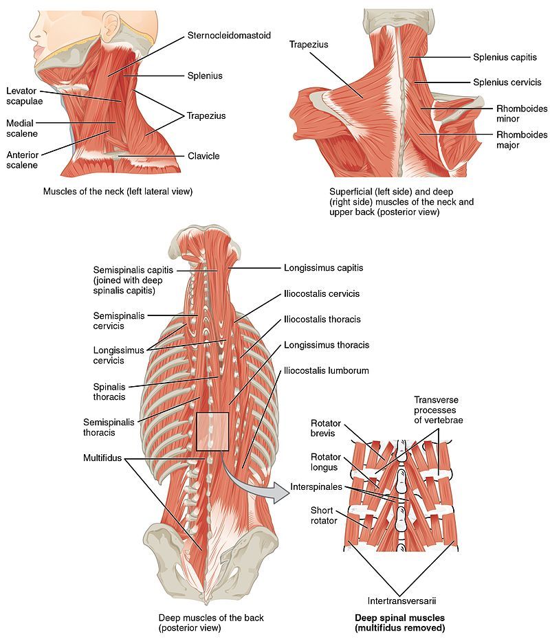
Just need a glimpse, leave your valuable advice let us know , and subscribe us! Deep back muscles diagram the superficial layer contains the splenius cervicis and splenius capitis muscles. For example, some muscles located in the chest also help move the shoulders. We are pleased to provide you with the picture named muscles of lower back diagram.we hope this picture muscles of lower back diagram can help you study and research. Pain log more pain mapping tools We think this is the most useful anatomy picture that you need. The part of the nerve that emerges out of the spine is called the nerve root. Five pairs of lumbar spinal nerves labeled l1 to l5 branch off your spinal cord and exit through small holes between the vertebrae. A heating pad or warm baths may provide temporary pain relief. Human muscle system, the muscles of the human body that work the skeletal system, that are under voluntary control, and that are concerned with movement, posture, and. This is a tutorial to quickly s. Facebook twitter google+ linkedin stumbleupon tumblr pinterest reddit vkontakte share via email print. The back muscles represented on an anatomical chart and on a schematic view of the origin and insertion of the proper muscles of the back (iliocostal muscle of.
The pelvis at the bottom of the back and the shoulders at the top of the back give the back. The back muscles represented on an anatomical chart and on a schematic view of the origin and insertion of the proper muscles of the back (iliocostal muscle of. Below you'll see diagrams along with the names of the back muscles that may be the cause of your pain. They are in fact different, but all three work together to support your spine and to help protect it from injury. The muscle elevates, depresses, rotates, and retracts the scapula, or shoulder blade.
Figure Muscles Of The Back Contributed Statpearls Ncbi Bookshelf from www.ncbi.nlm.nih.gov The back consists of the spine, spinal cord, muscles, ligaments, and nerves. The muscles, bones, ligaments, and tendons in the back can all be injured and cause back pain. These muscles include the large paired muscles in the lower back, called erector spinae, which help hold up the spine, and gluteal muscles. Related posts of muscles of the lower back and buttocks diagram body muscle structure. Build wide lats with this back building exercise. Three types of back muscles that help the spine function are extensors, flexors and obliques. Human muscle system, the muscles of the human body that work the skeletal system, that are under voluntary control, and that are concerned with movement, posture, and. Back to tracking tools main page.
Pain log more pain mapping tools
The intermediate layer contains the erector spinae muscles, whose many functions include the extension and lateral flexion of the spine, head and neck. The trapezius and latissimus dorsi muscles connect the upper limb to the vertebral column. Start with 5 to 10 minutes of moderate cardio to get your blood pumping and start to awaken your muscles. Deep back muscles diagram the superficial layer contains the splenius cervicis and splenius capitis muscles. The muscles of the back can be arranged into 3 categories based on their location: We hope this picture anatomy of back muscles diagram can help you study and research. Build wide lats with this back building exercise. This muscle is a major generator of lower back and hip pain, as well as being responsible for complaints of a burning sensation along the posterior superior iliac spine (psis) and sacroiliac joint. For example, some muscles located in the chest also help move the shoulders. Back pain due to muscle strain will usually get better on its own, but you can take steps to make yourself more comfortable. Postural and active movement muscle, used to tilt and turn the head and neck, shrug, steady the shoulders, and twist the arms. Superficial, intermediate, deep and deepest layers.these muscles lie on each side of the vertebral column, deep to the thoracolumbar fascia they span the entire length of the vertebral column, extending from the cranium to the pelvis Superficial back muscles, intermediate back muscles and intrinsic back muscles.the intrinsic muscles are named as such because their embryological development begins in the back, oppose to the superficial and intermediate back muscles which develop elsewhere and are therefore classed as extrinsic muscles.
Share :
Post a Comment
for "Back Muscles Diagram : Muscles Of The Trunk Anatomy Diagram Pictures Kenhub"


Post a Comment for "Back Muscles Diagram : Muscles Of The Trunk Anatomy Diagram Pictures Kenhub"